
Last month, I ended my week with yet another ankle surgery on a patient who sustained an “ankle sprain” three years ago. The patient was treated at an urgent care with Motrin and a brace. The patient was told the X-rays were negative for a fracture. About three weeks after the injury, the patient did some home ankle rehabilitation exercises and felt better. About a year after the injury, the patient complained of residual ankle pain and felt ankle stiffness especially in the morning. An MRI was ordered, which showed nothing wrong.
Often, an MRI can show detailed pathology in the ankle. But this is another example of a patient with a negative ankle MRI examination for ligament or cartilage damage, but with residual symptoms.
After a few months of physical therapy, the patient decided to proceed with a minimally invasive procedure called ankle arthroscopy.
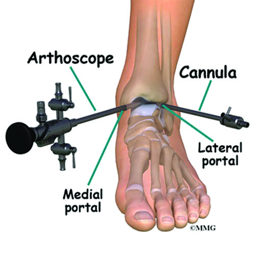
Ankle arthroscopy is a surgical procedure that uses a fiber-optic viewing camera and small surgical instruments to operate in and around the ankle joint through tiny incisions. Ankle arthroscopy is performed for the surgical evaluation and treatment of a variety of ankle conditions. Arthroscopy can also be performed at the knee, shoulder and hip joints.
Since 1984, ankle arthroscopy has become a widespread and versatile technique that many foot and ankle surgeons use to treat ankle pathology. Its popularity has grown because surgeons can perform the procedures quickly with increased accuracy and low complication rates. Arthroscopy also offers shorter recovery times in comparison to other operative techniques for treating ankle joint pathology. Over the last few decades, arthroscopy has become an advanced treatment method offering enhanced visualization of the joint and often a quick recovery for the patient.
Arthroscopy is very valuable when there is a build up of debris in the ankle from torn cartilage, torn ligaments, bone chips or spurs and/or thickened tissue that is impinging the joint. In particular, it allows unrivaled diagnostic visualization of the ankle joint with the opportunity to treat the pathology at the same time.We can now do this procedure in the office if the patient does not want any general anesthesia. A tiny camera at the tip of the needle allows the surgeon to evaluate the joint for pathology. Here at CMD, we use The Mi- Eye device.
Recently, Athrex, a well-known orthopedic device company developed the Nanoscope which allows both visual diagnostic evaluation of the joint as well as the ability to treat the pathology all in an office setting with the patient awake, or with only local anesthesia.
Thankfully, my patient did not want to “get by” with years of anti-inflammatory and pain medicines. The patient promptly chose to undergo ankle arthroscopy. It is important not to ignore residual ankle symptoms several years after an injury as it can lead to worse pain, impingement syndromes and ultimately arthritis.
Dr. Popowitz is a Board Certified Podiatrist and Foot & Ankle Surgeon. He is the chief of The Foot and Ankle Division at the Center for Musculoskeletal Disorders.Offices are located in Englewood, NJ, Flushing Queens, NY, and Brooklyn NY. To schedule an appointment or for more information please call 201.510.3777 or 718.509.1900.