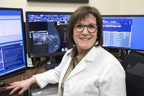
(Courtesy of Englewood Hospital) Growing up in a home surrounded by impressionist paintings, Mindy Goldfischer, MD, was taught by her parents to appreciate art. When looking at radiological studies in medical school, she found image interpretation similar to art interpretation, and she gravitated to the field of radiology.
Dr. Goldfischer has served as the chief of breast imaging at Englewood Health for more than 25 years. In this interview, she discusses what Jewish women should know about breast cancer screening.
At what age should Jewish women begin breast cancer screening?
The American College of Radiology (ACR) recommends that women with an average risk of developing breast cancer begin annual screening mammography at age 40. Because younger women tend to have dense breast tissue, which can obscure masses, annual mammograms are important; sometimes a supplemental breast ultrasound is indicated. High-risk women—those who have a first-degree relative (mother, sister, daughter) with breast cancer or a BRCA gene mutation—should begin annual screening 10 years earlier, but not before the age of 25.
Is there an upper age limit for mammography?
The ACR recommends that women have an annual mammogram from ages 40 to 75. After the age of 75, women should consult their physician to determine whether they need additional screening. Generally, someone with a life expectancy of five to 10 years should continue with screening mammography, possibly at less frequent intervals.
Who is at high risk?
Most women who develop breast cancer have no known risk factors. Only 5–10 percent of breast cancers occur in women with a genetic mutation, usually BRCA1 or BRC2.
Women with a history of breast cancer are at increased risk for another breast cancer.
Women with a genetic mutation or a first-degree relative with one. Women of Ashkenazi Jewish descent are at increased risk for BRCA mutation, as well as other less-common mutations, such as CHEK2.
Women with multiple family members (maternal or paternal), especially first-degree relatives, who have had breast cancer. The age at diagnosis is important, with premenopausal occurrence increasing risk.
Chest radiation before the age of 30 increases the risk of breast cancer (as well as cardiac disease).
Women who have had one or more breast biopsies for ADH (atypical ductal hyperplasia) or LCIS (lobular carcinoma in situ).
Should all Jewish women be tested for genetic mutations?
Only 10 percent of Ashkenazi Jewish women carry BRCA gene mutations, so it is important to consider all risk factors before proceeding with genetic testing. At Englewood Health, all women having a mammogram are seen by a nurse practitioner or physician’s assistant, who performs a breast exam and obtains a history, to assess the risk of breast cancer. We use the Tyrer Cuzick model, which calculates a person’s lifetime risk of developing breast cancer. A score of greater than 20 percent is considered high risk.
What are the advantages of 3D mammography?
At The Leslie Simon Breast Care and Cytodiagnosis Center at Englewood Health, almost all mammograms are 3D. With the traditional 2D mammogram, the X-ray tube is stationary and the breast tissue overlaps in the image. With a 3D mammogram, the X-ray tube moves in an arc around the breast. Images are obtained from multiple angles and synthesized by a computer, which creates thin slices that can be viewed individually. At Englewood Health, a special computer algorithm achieves 3D mammograms with the same radiation dose as 2D mammograms.
What are dense breasts, and why is ultrasound recommended for screening?
Dense breasts contain a larger percentage of glandular and connective tissue, which appears white on both 2D and 3D mammograms. After menopause, breasts often become less dense, as glandular tissue is replaced by fat, which appears gray or black on mammograms. Because cancers appear white on mammograms, a cancer is more easily discerned in a fatty breast than in a dense one. Ultrasound uses sound waves to visualize solid masses that can be obscured by breast tissue. Women with dense breast tissue may benefit by having an ultrasound in addition to their annual mammogram.
Does Englewood Hospital offer same-day mammogram results?
We offer all of our patients the option of remaining in the center while their mammograms are read. If additional mammogram views are needed, they are done the same day. If there is a suspicious finding on a mammogram and an ultrasound is recommended, the patient has the option of having it the same day. Many ultrasound-guided biopsies can also be performed during the same visit.
If diagnosed with breast cancer, what can a patient expect from the Englewood team?
Our Lefcourt Family Cancer Treatment and Wellness Center takes a team approach. A weekly multidisciplinary conference (tumor board) is held where all new breast cancer cases are presented and discussed. The conference is attended by pathologists, radiologists, medical and radiation oncologists, breast and plastic surgeons, the patient navigator, the advanced practice nurse certified in genetics, the clinical trials coordinator, and social worker. We review test results and then discuss the best approach to treatment, individualized according to each patient’s type and stage of breast cancer in accordance with national guidelines, as well as patients’ medical and social history. This team approach allows for specialists to consider multiple options in one setting to meet each patient’s individual needs. The patient’s progress is monitored at subsequent meetings while undergoing treatment, so additional recommendations can be made as needed. This conference has been held weekly for over 25 years. During that time, tremendous changes have occurred in the diagnosis and treatment of breast cancer and patients have benefited greatly from the dialogue and exchange of information that takes place in this forum.
Mindy Goldfischer, MD, is chief of breast imaging and a leader in The Leslie Simon Breast Care and Cytodiagnosis Center at Englewood Health.


